Looking for Pathology WSI Datasets? Here’s How Expert Medical Data Annotation Turns Raw Slides into AI-Ready Data


Shailesh Maurya is a Full-Stack Software Developer specializing in React, Node.js, and scalable backend solutions. He builds high-performance web applications with a strong focus on user experience, reliability, and clean architecture. He also has experience in RAG development, working with LLMs such as Ollama and ChatGPT, along with vector databases like PGVector to build intelligent AI-powered solutions.
Are you searching for pathology datasets, whole-slide image (WSI) datasets, histopathology datasets, tumor segmentation datasets, or annotated cancer datasets to train your AI model?
Finding medical images is only the first part of building a successful healthcare AI solution.
For an AI or machine learning model to understand a pathology slide, the data needs to be accurately annotated, segmented, classified, and quality-checked. Depending on the application, you may need tumor regions outlined, individual nuclei identified, tissue types classified, biomarkers labeled, or diagnostic regions marked.
This is where a specialized pathology data annotation service provider can help.
Srishta Technology Private Limited provides medical data annotation services to help AI companies, healthcare technology teams, researchers, and medical imaging organizations transform raw pathology data into structured, AI-ready datasets.
Why Raw Pathology Datasets Are Not Enough for AI
A whole-slide image may contain millions or even billions of pixels and a huge amount of visual information. But an AI model does not automatically understand which area contains tumor tissue, which cells are lymphocytes, where necrosis is present, or which regions are diagnostically important.
The model needs reliable ground truth.
For example, a raw WSI can be transformed into structured training data containing:
Tumor vs. Non-Tumor Regions → Tissue Classes → Cells & Nuclei → Regions of Interest → Biomarker Information → Diagnostic Labels
The quality of these annotations directly influences the quality of the dataset used for model development.
Who Provides Pathology Data Annotation?
A medical data annotation company provides trained annotation teams that convert raw medical images and related data into structured labels for AI and machine learning.
Pathology annotation is considerably more specialized than general image labeling.
Depending on the project, annotators may need to distinguish between tumor and normal tissue, identify nuclei, classify immune cells, locate mitotic figures, identify necrosis, evaluate staining patterns, or mark clinically relevant regions.
For complex projects, medical-domain expertise and expert review can therefore become important parts of the annotation workflow.
Pathology and Whole-Slide Image Annotation Services
Srishta Technology can support different levels of pathology annotation based on the client's taxonomy, annotation guidelines, model requirements, and expected ground truth.
1. Whole-Slide Image (WSI) Annotation
Whole-slide images are high-resolution digital representations of pathology slides.
WSI annotation can involve marking relevant regions across a slide and assigning labels that help machine learning models identify important pathological patterns.
Typical WSI annotation requirements can include:
- Tumor vs. non-tumor regions
- Tumor segmentation
- Tissue segmentation
- Cell and nuclei annotation
- Necrosis annotation
- Mitotic figures
- Immune-cell annotation
- Biomarker/IHC annotation
- ROI marking
- Cancer grading or staging labels
- Slide-level diagnosis
- Pixel-level segmentation masks
The exact annotation approach should be defined according to the intended AI application.
2. Tumor vs. Non-Tumor Annotation
One of the most common requirements in computational pathology is teaching an AI model to differentiate tumor tissue from non-tumor tissue.
Annotators can outline relevant regions according to project guidelines and classify them as:
Tumor | Non-Tumor | Normal Tissue | Other Defined Tissue Classes
These annotations can be used to develop models for tumor detection, localization, classification, and segmentation.
3. Tumor Segmentation
Tumor segmentation provides more detailed information than simply assigning a tumor label to an entire image.
The objective is to identify the location and boundaries of tumor regions.
Depending on the project, annotations can be created as polygons, regions, or pixel-level segmentation masks.
This provides the AI model with precise spatial information about where tumor tissue occurs within the pathology image.
4. Cell and Nuclei Detection, Segmentation & Classification
Cellular-level analysis is another important area of computational pathology.
Depending on the use case, annotation can include:
- Nuclei detection
- Nuclei segmentation
- Nuclei classification
- Cell detection
- Cell segmentation
- Cell classification
Individual cells can also be assigned to predefined categories based on the project's taxonomy.
For example:
Tumor Cells | Lymphocytes | Plasma Cells | Macrophages | Other Cells
This type of annotation can support AI applications involving cellular morphology, density, distribution, and the tumor microenvironment.
5. Tissue Segmentation and Classification
A pathology slide can contain several different types of tissue.
Tissue segmentation involves dividing the image into meaningful regions and assigning each region to a specific tissue category.
Typical classes can include:
- Tumor
- Stroma
- Necrosis
- Normal tissue
- Background
- Other client-defined tissue categories
Accurate tissue segmentation helps AI systems understand both the type and spatial distribution of tissue structures.
6. Cancer Grading and Staging Labels
Some pathology AI projects require labels related to cancer grade or stage.
These tasks require clearly defined clinical guidelines and the appropriate level of medical expertise.
Depending on project requirements, qualified experts can be incorporated into the review and adjudication process to establish reliable ground truth.
7. Mitotic Figure Annotation
Mitotic figures can be important indicators in the assessment of several cancers.
Annotation may involve identifying individual mitotic figures using:
- Point annotation
- Bounding boxes
- Classification labels
- Region-specific annotations
Because mitotic figures can be challenging to distinguish, appropriate expert review can be incorporated when required.
8. Necrosis Annotation
Necrotic tissue can be annotated separately from viable tumor, normal tissue, and other surrounding structures.
Depending on the AI model requirements, necrosis can be represented through:
Region Annotation | Polygon Annotation | Tissue Classification | Segmentation Masks
This helps create more detailed tissue-level ground truth.
9. Lymphocyte and Immune-Cell Annotation
The tumor microenvironment contains different immune-cell populations that may be relevant to computational pathology research.
Annotation requirements may include identification or classification of:
- Lymphocytes
- Plasma cells
- Macrophages
- Tumor cells
- Other immune-cell categories
The classes can be customized according to the client's research objectives and annotation guidelines.
10. Biomarker and IHC Annotation
Immunohistochemistry (IHC) annotation can help prepare structured datasets for AI models designed to analyze biomarker expression.
Depending on the staining method, biomarker, and project guidelines, annotation may include:
- Positive cells
- Negative cells
- Staining intensity
- Biomarker-positive regions
- Cell-level classifications
- Tissue-level staining regions
The labeling criteria should be established during project calibration to ensure consistency across the dataset.
11. ROI – Region of Interest Annotation
Not every part of a pathology slide has equal diagnostic or analytical importance.
Region of interest (ROI) annotation identifies areas that are particularly relevant to the project.
These may include:
- Suspected tumor areas
- Representative tissue regions
- High-cell-density regions
- Regions containing important morphological features
- Areas requiring detailed cellular annotation
ROI annotation can also help make WSI projects more efficient by focusing detailed labeling on relevant areas.
12. Pathology Report & NLP Annotation
Pathology AI does not always involve images alone.
Pathology reports contain valuable unstructured medical information that can be transformed into structured datasets for natural language processing (NLP) applications.
Depending on project requirements, annotation can cover information such as:
- Diagnosis
- Tumor type
- Anatomical site
- Histological findings
- Grade
- Stage
- Biomarker information
- Medical entities
- Relationships between clinical entities
This can support medical NLP and multimodal AI systems that combine image and text information.
13. Slide-Level Diagnosis Labels
Not every machine learning project requires detailed pixel-level annotation.
For certain classification applications, the complete slide may receive a slide-level diagnosis or classification label.
For example:
Whole-Slide Image → Tumor
Whole-Slide Image → Non-Tumor
Or the slide can be assigned to a more specific diagnostic category according to the project's defined ground truth.
14. Pixel-Level Segmentation Masks
When precise localization is required, pixel-level segmentation masks can be created.
This approach identifies the exact image regions corresponding to a particular structure or class.
Pixel-level masks may be required for:
- Tumor segmentation
- Tissue segmentation
- Nuclei segmentation
- Cell segmentation
- Necrosis segmentation
- Biomarker-positive regions
These detailed annotations are particularly useful for computer vision models designed for segmentation tasks.
Why Pathologist-Reviewed Ground Truth Matters
Medical AI requires high-quality reference data.
For projects requiring clinical ground truth, a review workflow can incorporate qualified medical experts or pathologists according to the complexity and requirements of the project.
Top Data Labeling Companies in United States 2026
A typical workflow may look like:
Initial Annotation → Medical Review → Quality Check → Disagreement Resolution → Expert Adjudication → Final Ground Truth
This process helps improve consistency and provides stronger reference data for model training, validation, and testing.
Why Choose Srishta Technology for Pathology Data Annotation?
Choosing the right medical data annotation company is not simply about finding someone who can draw polygons or create segmentation masks.
Medical AI projects require accuracy, domain understanding, consistent guideline implementation, scalable annotation workflows, and strong quality assurance.
Here is how Srishta Technology can support pathology AI projects.
Medical-Domain Expertise
Srishta Technology can create project teams based on the level of medical expertise required for a particular annotation task.
For projects involving complex medical terminology or clinical interpretation, appropriately trained medical professionals can be incorporated into the workflow.
Customized Annotation Taxonomy
Every pathology dataset is different.
Instead of using a fixed annotation structure, Srishta Technology can work according to your specific taxonomy.
For example:
Tissue Classification
Tumor | Stroma | Necrosis | Normal Tissue
Cell Classification
Tumor Cell | Lymphocyte | Plasma Cell | Macrophage
The annotation workflow can be customized according to your classes, attributes, quality criteria, and expected output.
Pilot Annotation Before Full Production
For large medical annotation projects, we recommend starting with a pilot.
Our workflow can follow:
Requirement Understanding → Taxonomy → Pilot Annotation → Client Calibration → Production Annotation → Multi-Level QA → Expert Review → Delivery
The pilot allows both teams to align on guidelines and expected accuracy before scaling annotation across the complete dataset.
Multi-Level Quality Assurance
Quality control can be designed according to the complexity of the project.
Depending on requirements, the workflow may include:
- Annotator self-review
- Peer review
- Dedicated QA
- Medical review
- Guideline compliance checks
- Expert adjudication
The goal is to deliver consistent and reliable annotations suitable for downstream AI development.
From Raw WSI to AI-Ready Pathology Dataset
The process of transforming raw pathology images into structured AI training data can be summarized as:
Raw Whole-Slide Images
↓
ROI Identification
↓
Tumor & Tissue Segmentation
↓
Cell/Nuclei Annotation
↓
Classification & Clinical Labels
↓
Medical/Pathologist Review
↓
Quality Assurance
↓
Final Ground-Truth Dataset
↓
AI Model Training, Validation & Testing
This is how raw medical images become structured and meaningful training data for computational pathology applications.
Looking for a Pathology Data Annotation Company in India?
If you already have pathology images but need high-quality medical data annotation, Srishta Technology can help transform your raw data into structured datasets according to your AI model requirements.
Before starting annotation, we recommend defining:
- Type of pathology data
- Number of slides/images
- Cancer or tissue type
- Required annotation classes
- Slide-, ROI-, cell-, or pixel-level requirements
- Annotation guidelines
- Expected output format
- Required medical expertise
- Pathologist review requirements
- Quality and accuracy expectations
Once the requirements are clear, a small pilot project can be completed to establish quality expectations before moving to full-scale production.
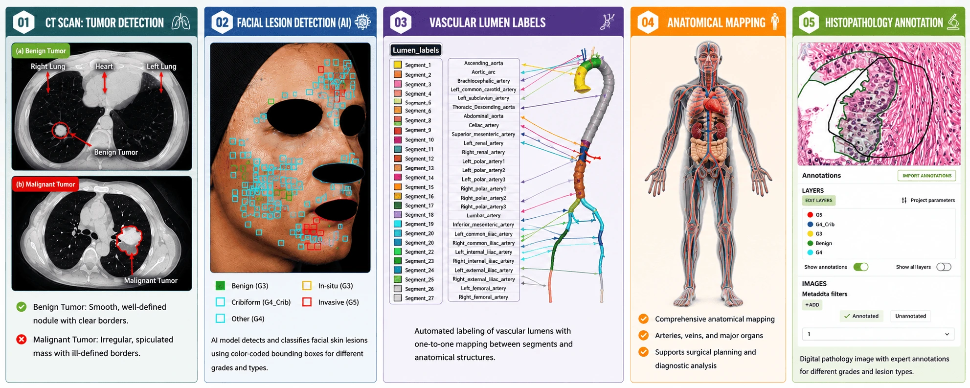
Frequently Asked Questions About Pathology Data Annotation
What is pathology data annotation?
Pathology data annotation is the process of labeling digital pathology images or related medical information to create structured ground truth for AI and machine learning models. It can include tumor segmentation, tissue classification, nuclei detection, cell classification, ROI marking, biomarker annotation, and diagnostic labels.
What is WSI annotation?
WSI annotation means labeling whole-slide images used in digital pathology. Depending on the project, annotations may identify tumors, tissues, nuclei, cells, necrosis, mitotic figures, immune cells, biomarkers, and diagnostically relevant regions.
What are the typical WSI annotation requirements?
Typical requirements include tumor segmentation, tumor vs. non-tumor classification, nuclei detection and segmentation, tissue classification, cell classification, ROI annotation, mitotic figure annotation, IHC annotation, grading/staging labels, and slide-level diagnosis.
Can Srishta Technology provide tumor segmentation annotation?
Yes. Srishta Technology can support tumor segmentation according to client-defined annotation guidelines, including outlining tumor and non-tumor regions and creating region- or pixel-level annotations.
Can you annotate cells and nuclei?
Yes. Annotation requirements can include cell/nuclei detection, segmentation, and classification according to the classes defined for the project.
Can you annotate lymphocytes and immune cells?
Yes. Depending on project requirements and image characteristics, annotation can include lymphocytes, plasma cells, macrophages, tumor cells, and other specified immune-cell classes.
Do you provide IHC and biomarker annotation?
Yes. IHC annotation can include positive and negative cell labels, staining intensity, biomarker-positive regions, ROI annotation, and other project-specific classifications.
Can you create pixel-level segmentation masks?
Yes. Pixel-level masks can be created for suitable tumor, tissue, nuclei, cell, necrosis, and biomarker segmentation requirements.
Can you annotate pathology reports for NLP?
Yes. Pathology report annotation can support medical NLP projects through the labeling of diagnoses, tumor types, anatomical sites, biomarkers, grades, stages, and other client-defined medical entities.
What is pathologist-reviewed ground truth?
Pathologist-reviewed ground truth refers to annotations or diagnostic labels that have been reviewed or adjudicated by qualified pathology experts according to a defined project protocol. This provides stronger reference data for medical AI development and evaluation.
Why should medical AI companies use specialized annotation providers?
Medical images can contain complex clinical information that requires specialized knowledge. A medical data annotation provider can combine annotation processes, domain expertise, quality assurance, and expert review to create more reliable AI training datasets.
How do I start a pathology annotation project with Srishta Technology?
Share your sample pathology data, annotation guidelines, taxonomy, expected output format, approximate volume, and expert-review requirements.
Srishta Technology can evaluate the requirements and begin with a small pilot annotation.
Once the annotation quality and guidelines are approved, the workflow can be scaled for production.
Build Better Pathology AI with High-Quality Ground Truth
If you are searching for pathology datasets for AI, remember that acquiring images is only one part of the process.
The real value comes from transforming those images into accurately annotated, structured, quality-checked, and AI-ready ground truth.
From whole-slide image annotation and tumor segmentation to nuclei detection, tissue segmentation, mitotic figures, immune-cell annotation, IHC annotation, ROI marking, pathology NLP, slide-level diagnosis, and pixel-level segmentation, Srishta Technology can support different stages of pathology dataset preparation.
Looking for a reliable pathology and medical data annotation partner?
Start with a small pilot annotation with Srishta Technology Private Limited. Evaluate the quality and accuracy first, align the workflow with your requirements, and then scale your pathology annotation project with confidence.
 +91 93543 34258
  +91 93543 34258